What does a lameness examination on your horse entail?
Lameness is one of the most common reasons our equine veterinarians are called to look at your horse.
It can be a significant cause of pain or distress and may even be life threatening. Meanwhile, more subtle lameness can be performance-limiting, causing gait irregularities, affecting speed, or contributing to behavioural abnormalities.
Fully investigating lameness in your horse requires some detective work, involving a combination of:
a full clinical examination,
palpation of the limbs from nose to tail and top to toe,
examination during walking and trotting in a straight line, while being lunged, and often during exercise (including being ridden).
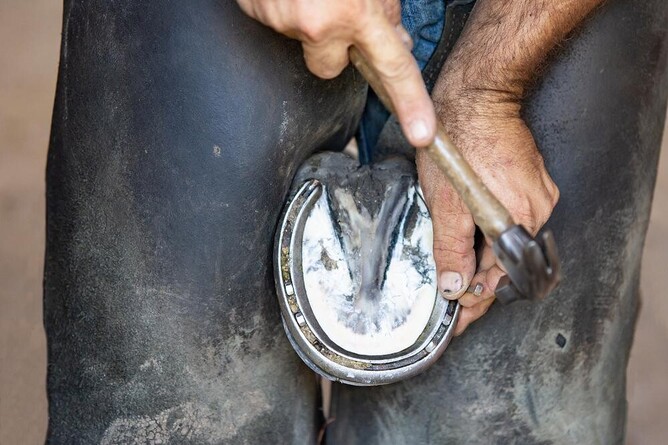
On occasions, the source of pain will be obvious, e.g. pain on hoof testers representing a hoof abscess, wound, or fractured pedal bone; or acute lameness, following exercise, where there is a marked painful response on palpation (with high suspicions of a fracture).
However, often the cause of lameness may be more insidious in onset, where multiple areas of pain may be appreciated (that don’t always cause the lameness), or alternatively the horse doesn't respond to any areas of pain when palpated.
In these cases, the use of local, regional anaesthesia (nerve blocks), intra-articular anaesthesia (joint blocks), or intrasynovial anaesthesia (tendon sheath blocks) are used to ‘numb’ the region of interest.
By reducing or numbing the pain, lameness is effectively resolved, helping the clinician to localise the lameness to a general region or specific joint.
The use of imaging
Following localisation of the lameness, our vets will then look at using imaging modalities to determine the nature of the problem, it's cause, and to guide the prognosis.
Such imaging modalities would normally include radiographs (X-rays) and the use of ultrasound. In general, X-rays can be used to detect boney changes (fractures, arthritis, bone cysts, bone infections etc), while ultrasound is often used to detect soft tissue abnormalities (tendon & ligament injuries, soft tissue damage within and around joints, or muscle damage etc).
In some horses, referral to Christchurch for a bone scan (scintigraphy) may be recommended. This involves the injection of a radioactive isotope into the vein, before the horse is placed in front of a camera which detects the ‘counts’. Areas of increased inflammation will have higher counts and, therefore, be identified as a ‘hotspot’.
Such horses recommended for a scintigraphy may be unresponsive to nerve blocks or become difficult to handle with needle placement.
On occasions where an upper limb lameness is suspected, or a lower limb lameness cannot be identified radiographically or on ultrasound, a bone scan is also recommended.
Computed tomography (CT) and magnetic resonance imaging (MRI) is also used in other areas of New Zealand and the world to aid lameness investigation.
Commonly asked questions
How long does a lameness investigation take?
This can vary depending on the severity of the lameness or how acute it is. Lower limb fractures may be quickly diagnosed, but more in depth lameness work-ups may take several hours, and sometimes carry over into the next day.
How much does it cost?
Costs can vary substantially based on the diagnostics and treatment plan required. Diagnostics can include radiographs, nerve blocks or even referral for a bone scan. Please contact the clinic to discuss your horse's needs.
Where can a lameness examination be performed?
We can perform a lameness investigation either on-site at VetSouth Invercargill, or remotely at a suitable location that has a safe, clean, flat firm footing (e.g. tarseal). An arena or track is recommended if exercising your horse is required.
Most of our equipment is portable, so provided you have a power source, we are able to complete a large number of diagnostics off site.
My horse is lame, should I still work it until my veterinary appointment?
With acute severe lameness, particularly following fast work or jumping/cross country, work should be stopped, the lower leg supported and the horse immediately taken to a stable. These are veterinary emergencies and veterinary attention should be sought immediately.
Acute fractures of the pastern, cannon bone, pedal bone or knee (carpus) are the most common fractures that we see in racehorses, particularly after fast work, however fractures in any bone are not uncommon in horses in the paddock.
Obvious lamenesses where it is easily identified at a trot should be rested and an appointment made straight away.
Some of these injuries may be stress fractures or soft tissue injuries (e.g. tendon injuries) that require assessment and treatment before a horse is turned out.
Subtle intermittent lameness that comes and goes can be frustrating to assess, as resting the horse for 2-3 days may result in the lameness disappearing, and these horses often show very little lameness when they are examined.
Discussion of individual cases with the veterinarian at the time the appointment is made is recommended to allow these cases to be investigated fully.
For any further questions regarding lameness investigations in your horse, or for recommendations on management and treatment, please feel free to contact us at VetSouth Equine.